Related Projects
- Quantification of Geographic Atrophy in Fundus Autofluorescence Images for Diagnosis of Age-Related Macular Degeneration
- Interactive Segmentation for Geographic Atrophy in Retinal Fundus Images
- Retinal Vessel Segmentation using Multi-Scale Wavelet Frame Analysis
- Coarse to Fine Segmentation of Stargardt Rings using an Expert Guided Dual Ellipse Model
- Bayesian Transductive Markov Random Fields for Interactive Segmentation in Retinal Disorders
- A Software Tool for Retinal Image Analysis (RIALAB)
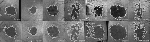
Quantification of Geographic Atrophy in Fundus Autofluorescence Images for Diagnosis of Age-Related Macular Degeneration
Fundus auto-fluorescence (FAF) images with hypo-fluorescence indicate geographic atrophy (GA) of the retinal pigment epithelium (RPE) in age-related macular degeneration (AMD). Manual quantification of GA is time consuming and prone to inter- and intra-observer variability. Automatic quantification is important for determining disease progression and facilitating clinical diagnosis of AMD. In this paper we describe a hybrid segmentation method for GA quantification by identifying hypo-fluorescent GA regions from other interfering retinal vessel structures. First, we employ background illumination correction exploiting a non-linear adaptive smoothing operator. Then, we use the level set framework to perform segmentation of hypo-fluorescent areas. Finally, we present an energy function combining morphological scale-space analysis with a geometric model-based approach to perform segmentation refinement of false positive hypo- fluorescent areas due to interfering retinal structures. The clinically apparent areas of hypo-fluorescence were drawn by an expert grader and compared on a pixel by pixel basis to our segmentation results. The mean sensitivity and specificity of the ROC analysis were 0.89 and 0.98%.
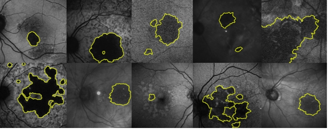
Interactive Segmentation for Geographic Atrophy in Retinal Fundus Images
Fundus auto-fluorescence (FAF) imaging is a non-invasive technique for in vivo ophthalmoscopic inspection of age-related macular degeneration (AMD), the most common cause of blindness in developed countries. Geographic atrophy (GA) is an advanced form of AMD and accounts for 12-21% of severe visual loss in this disorder [3]. Automatic quantification of GA is important for determining disease progression and facilitating clinical diagnosis of AMD. The problem of automatic segmentation of pathological images still remains an unsolved problem. In this paper we leverage the watershed transform and generalized non-linear gradient operators for interactive segmentation and present an intuitive and simple approach for geographic atrophy segmentation. We compare our approach with the state of the art random walker [5] algorithm for interactive segmentation using ROC statistics. Quantitative evaluation experiments on 100 FAF images show a mean sensitivity / specificity of 98.3 / 97.7% for our approach and a mean sensitivity / specificity of 88.2 / 96.6% for the random walker algorithm.
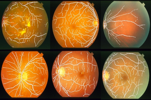
Retinal Vessel Segmentation using Multi-Scale Wavelet Frame Analysis
Fundus photography is a non-invasive technique for in vivo ophthalmoscopic inspection of retinal disorders. Quantitative information about the vascular network can facilitate clinical diagnosis of retinal diseases. We propose an algorithm for segmentation of the vascular network by using multi-scale analysis in selected wavelet channel frames.
We use the STAR database for testing our segmentation algorithm consisting of twenty datasets. The images are captured by a TopCon TRV-50 fundus camera with 35 degree field of view. An over-complete wavelet frame expansion is performed. We perform selective channel rejection in the decomposition tree followed by wavelet shrinkage and enhancement operators to separate retinal objects from background. The obtained vessel likelihood map is the basis for a Bayesian classifier into vessel and non-vessel employing shape, topology, and intensity cues.
The segmentations were compared to the expert grading on a pixel-by-pixel basis. The mean sensitivity and specificity were 0.88 ± 0.09 and 0.98 ± 0.02.
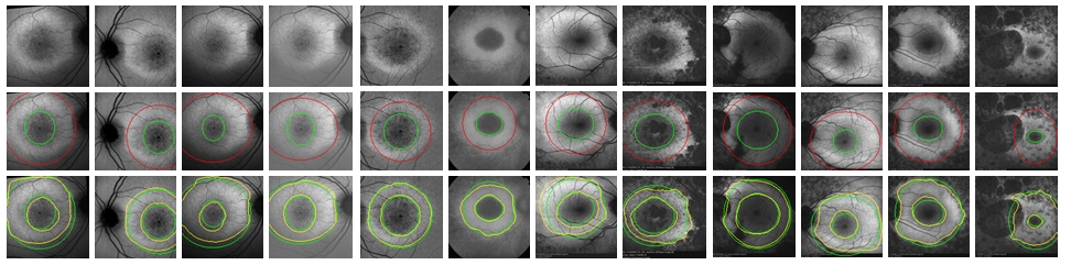
Coarse to Fine Segmentation of Stargardt Rings using an Expert Guided Dual Ellipse Model
Computer aided diagnosis in the medical image domain requires adaptive knowledge-based models to handle uncertainty, ambiguity, and noise. We propose an expert guided coupled dual ellipse model in a coarse to fine energy minimization framework. In our approach we enforce subspace model constraints by fusing domain knowledge and model information to guide the segmentation process on the fly. We apply our method to the task of retinal Stargardt segmentation a disease that manifests itself in a ring like structure around the macula. Quantitative evaluations on synthetic and real data sets show the performance of our framework. Experimental results demonstrate that our framework performance well with an area under the ROC curve of 0.93.
Bayesian Transductive Markov Random Fields for Interactive Segmentation in Retinal Disorder
A Software Tool for Retinal Image Analysis (RIALAB)
Related Publications
Noah Lee,Andrew F Laine,R. Smith, Coarse to Fine Segmentation of Stargardt Rings using an Expert Guided Dual Ellipse Model Proceeding of 30th Annual International Conference of the IEEE Engineering in Medicine and Biology Society (EMBC), pp. 2250-2253, Vancouver, Canada, 2008
Noah Lee,R. Smith,Andrew F Laine, Learning Non-Homogenous Textures And The Unlearning Problem With Application To Drusen Detection In Retinal Images Proceeding of 5th IEEE International Symposium on Biomedical Imaging (ISBI): From Nano to Macro, pp. 1215-1218, 2008
Noah Lee,Andrew F Laine, A hybrid segmentation approach for geographic atrophy in fundus auto-fluorescence images for diagnosis of age-related macular degeneration. 29th Annual International Conference of the IEEE, Engineering in Medicine and Biology Society (EMBS), 2007
M. Busuoic,Noah Lee,Andrew F Laine,R. Smith, Retinal vessel segmentation using multi-scale wavelet frame analysis The Association for Research in Vision and Ophthalmology (ARVO), Fort Lauderdale, Florida, USA, May 6-10, 2007
I. Barbazetto,M. Busuoic,Noah Lee,R. Smith,Andrew F Laine, Levelset Segmentation of Geographic Atrophy in Macular Autofluorescence Images The Association for Research in Vision and Ophthalmology (ARVO), Fort Lauderdale, Florida, USA, April 30-May 4, 2006
Noah Lee,R. Smith,Andrew F Laine, Interactive Segmentation for Geographic Atrophy in Retinal Fundus Images Proceedings of Asilomar Conference on Signals, Systems and Computers, IEEE Signal Processing Society, pp. 655-658, Pacific Grove, CA, USA, 2008
N. Gomes,M. Busuioc,R. Smith,Noah Lee,Andrew F Laine, Autofluorescence Image Analysis in Age-related Macular Degeneration (AMD) and Stargardt Disease (STGD) IEEE Signal Processing Society, Asilomar Conference on Signals, Systems and Computers, pp. 651 - 654, Pacific Grove, CA, USA, 2008