Active Projects
|
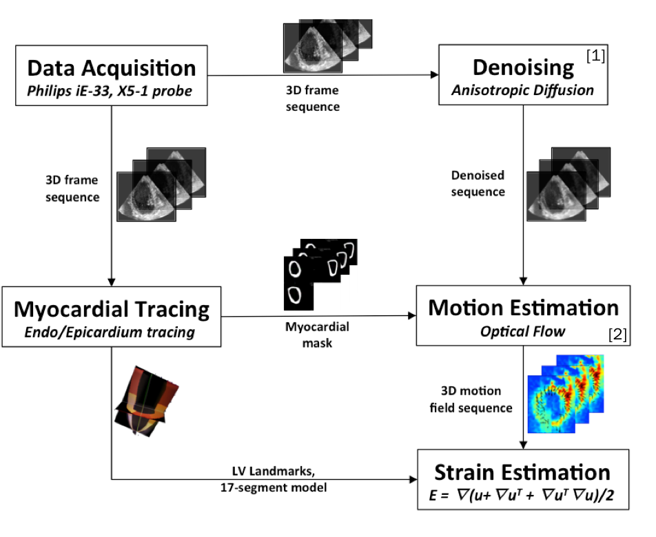
Clinical validation of cardiac strain measures with real-time 4D ultrasound (2008-2012) Period: 9/1/08 - current Short Info : This is a development of comparative studies of strain using cardiac MRI and 4D cardiac ultrasound. 4D optical flow based motion tracking algorithm is developed to extract fully 4D dynamic cardiac metrices. This is the first validation study of optical flow based strain estimation for 4D ultrasound with direct comparison based on in vivo data. more |
 |
|
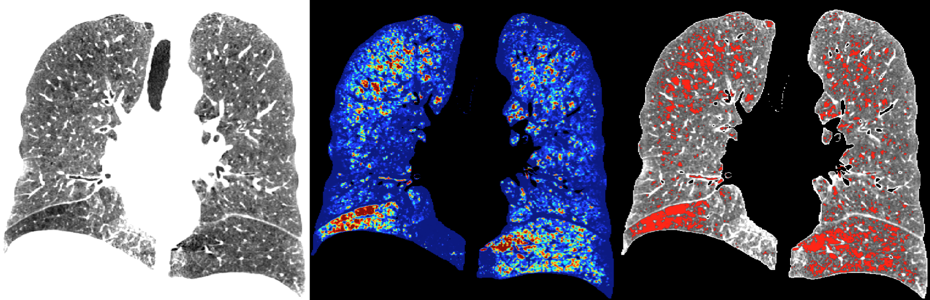
Adaptive quantification and subtyping of pulmonary emphysema from CT images Funded by National Institutes of Health (NHLBI)1 R01 Period: 2012 -current Short Info : Pulmonary emphysema is a condition involving alveolar wall destruction, contributing to chronic airflow limitation characteristic of chronic obstructive pulmonary disease (COPD). The processes underlying COPD are currently not well understood, even though the disease is a leading cause of morbidity and mortality worldwide. more |
 |
|
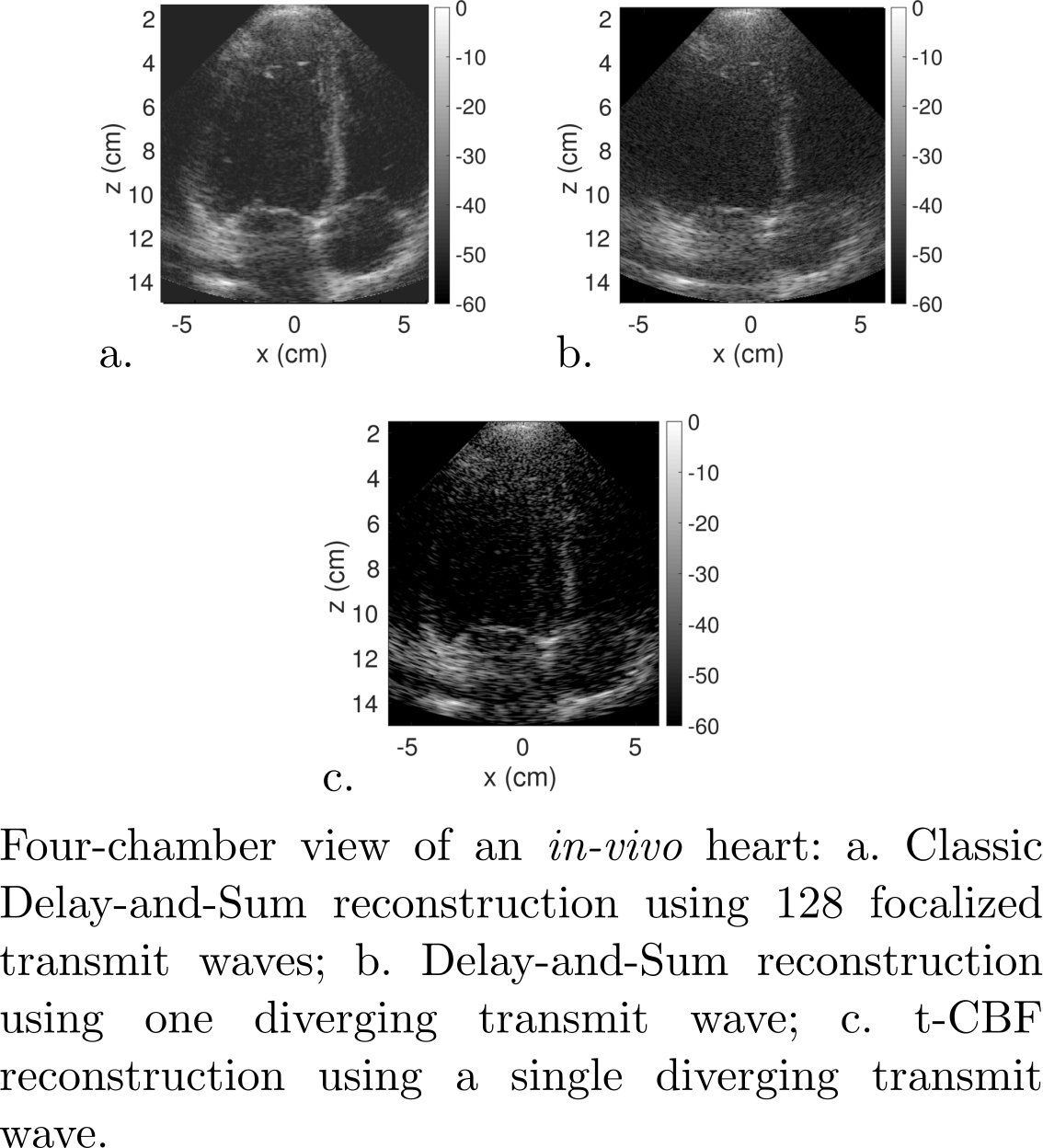
Time domain Compressive Beamforming (t-CBF): Application to echocardiography Period: 2011-current Collaboration with Philips Research North America Short Info: Ultrasound (US) imaging has been used in the medical field for over 20 years for applications ranging from fetus monitoring to tumor ablation, hence spanning both the diagnostic and therapeutic worlds. Its versatility and cost-effectiveness with respect to other imaging modalities such as MRI and X-ray make it a great tool in the medical imaging arsenal. However in spite of recent developments and enhancements, US imaging still has several drawbacks. In order to compute a single image, a vast amount of data has to be acquired and processed by the scanner impacting the frame rate of the machine among other aspects.
Independently, an inverse problem technique, Compressive Sensing (CS), has emerged and gained a lot of interest from researchers over the past decade because of its ability to recover undersampled information under some mathematical assumptions. After being thoroughly developed and supported as a mathematical theory, it was successfully applied to MRI, effectively decreasing the acquisition time significantly. Naturally, researchers from other fields have been trying to adapt CS to their own problems.
This work aims to adapt CS to US imaging and to develop a method that decreases the amount of data acquired while maintaining the image quality, relying on physical models, wavelets, and processing power to fill-in the gaps, so to speak.
|
 |
PAST PROJECTS
|
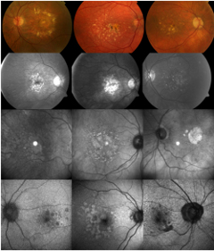
Quantitative Analysis of Retinal Images via Fluoroscopy (2005-2010) Period: 9/15/05 - 08/31/10 Short Info : Create robust and minimal intervention algorithms for segmentation and quantitation of the key abnormalities in retinal disorders: drusen, GA, hyper- and hypopigmentation in color photographs, hyper/hypofluorescent lesions in AF images, and hyper/hyporeflectance in IR images. Define disease metrics based on data fusion from the images in registration. more |
 |
|
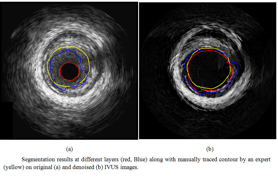
IVUS Image Analysis (2008-2009) Period: 9/1/08 – 8/31/09Collaboration with Dr. Bernhard Sturm, Volcano Rancho Cordova, Dr. Elsa D. Angelini, Telecom ParisTech, Paris, FranceShort Info: Design and implement on IVUS image denoising and lumen border detection. more |
 |
|
Automated detection of Protein Crystal Images Funded by National Institutes of Health (NESG) Period: 2000-2009 Short Info : High-throughput experiments with varying crystallization conditions are currently performed with the hopes that one or more conditions will provide leads for actual protein crystallization. The High-throughput experiments generate millions of images that then have to be analyzed. We propose an automated classification algorithm that can replace the need for crystallographers to manually classify these images. more |
 |
|
Development of non-Fourier MR Spectroscopy Protocols for Brain Imaging (2007-2012) Period: 8/1/2007 to 7/31/2012 Collaboration with Dr. Oded Gonen, Dr. Matilde Ingelese, Center for Biomedical Imaging of New York University’s Department of Radiology Short Info: The goal of this project is to develop acquisition and reconstruction methods of non-Fourier data for diagnosis of various diseases. more |
 |
|
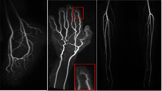
Non-Contrast Enhanced Magnetic Resonance Angiography Funded by NIH grant HL092439 Collaboration with Dr. Vivian Lee from the Center for Biomedical Imaging of New York University Department of Radiology Short Info: The objective of this project is to develop and test non-contrast-enhanced MRA for the diagnosis of patients with peripheral arterial disease at 3T. more |
 |
|
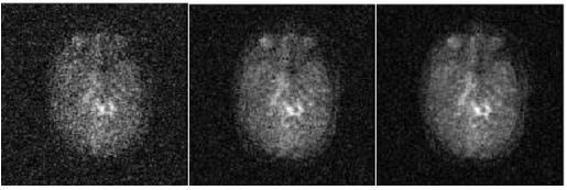
Non-invasive quantification of Positron Emission Tomography (PET) data Period: 2011-2014 Collaboration with Dr. Ramin Parsey from the Department of Psychiatry, Stony Brook University and Dr. John Mann from the Division of Molecular Imaging and Neuropathology, New York State Psychiatric Institute. Short Info: The goal of this project non-invasively determine the changes in blood levels of PET compound over time during a brain scan. more |
 |
|
Multi-modality analysis of dementia that combines shape analysis and funtional data Period: 2011-2014 Short Info: Develop a multi-modality framework for assessing Alzheimer’s disease utilizing brain region shape analysis and functional imaging more |
 |
|
Developing algorithms and techniques to measure structural and functional changes in the adult brain, for Alzheimer Disease Period: 2010-2015, Collaboration with the Taub Institute in the Department of Neurology Short Info: Khan UA, Liu L, Provenzano FA, Berman DE, Profaci CP, Sloan R, Mayeux R, Duff KE, Small SA. “Molecular drivers and cortical spread of lateral entorhinal cortex dysfunction in preclinical Alzheimer’s disease.” Nat Neurosci. 2013 Dec 22. |
|