Principal Investigator
Andrew F. Laine, D.Sc.
Students/Research Scientists
Elsa Angelini, PhD.
Viktor Gamarnik, MD.-PhD
Collaborators
Shunichi Homma, M.D., Department of Medicine, Columbia University
Marco R. DiTullio, M.D., Department of Medicine, Columbia University
Cesare Russo, M.D., Department of Medicine, Columbia University
Edward Lee, M.D., Department of Medicine, Columbia University
Jeffrey Holmes, M.D., Ph.D., Department of Biomedical Engineering and Medicine, University of Virginia
Katherine M. Parker, Department of Biomedical Engineering and Medicine, University of Virginia
Summary
Cardiac imaging technology has advanced dramatically in recent years. Specifically, Real-Time Three-dimensional (or RT3D, 4D) echocardiography is becoming increasingly attractive because of its ability to acquire full three-dimensional images of the heart over a full cardiac cycle within a few seconds. The complex 3D wall motion and temporal information contained in these four-dimensional (3D + time) data sequences has the potential to greatly enhance clinical diagnoses of the heart. However, most cardiac examination centers still depend on 2D echocardiography, and the temporal information in 4D cardiac imaging is often overlooked. Although tissue Doppler imaging (TDI) and strain imaging are widely used in 2D echocardiography, there is no counterpart for 4D echocardiography available clinically. This is because effective 4D quantitative analysis tools are not widely available. Without such tools, the voluminous information contained in one 4D image acquisition sequence remains prohibitive to analyze for routine clinical use. Metrics of cardiac dynamics, including displacements and strain measurements have been shown to be reliable and efficient indicators of cardiac abnormalities, such as regional ischemia and dyssynchrony.
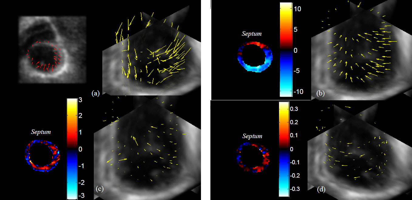
Figure 1: Example results in a heart transplant patient of metrics of cardiac dynamics: (a) displacement field; (b) radial displacement (in mm); (c) thickening (in 100%); (d) twist (in radian per mm).
Although echo-based techniques, such as TDI, strain imaging, and speckle tracking imaging (STI), can effectively measure these metrics in 2D, 4D cardiac motion and strain measurements have been mainly restricted to MR-based tissue tagging methods which offer non-invasive quantification of cardiac deformation. With recent developments in imaging techniques, post-processing methods, and validation studies, MR tagging has become a gold standard for motion and strain measurements in vivo. However, MR-based imaging techniques cannot be applied to all cardiac patients, such as patients with pace makers or those sensitive to long acquisition times in confined space, or the use of complicated exercise stress testing for assessing myocardium at risk. Thus 4D motion and strain measurements with MRI for these patient populations are not possible and remain problematic.
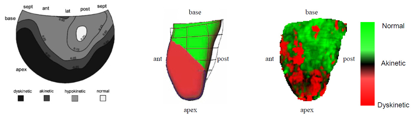
Figure 2: Wall motion abnormalities during finite element simulation of a proximal LAD occlusion: (a) Hammer map of corresponding endocardium with shading representing: normal wall motion (white), hypokinesis (light gray), akinesis (dark gray), and dyskinesis (black); (b) anterior view of the finite element model with a large simulated LAD occlusion.
Building on the last 8 years of collaboration with clinicians (Echo Cardiography Laboratory directed by Dr. Homma, Department of Medicine, Columbia University) and cardiac biomechanicians (Cardiac Biomechanics Group directed by Drs. Holmes and Costa, Department of Biomedical Engineering, Columbia University), our biomedical imaging group has recently developed an optical flow-based motion tracking algorithm for 4D echocardiography. Our method has been quantitatively validated for tracking the endocardium and the epicardium against manual tracing. We have evidence to suggest that several metrics of cardiac dynamics, including deformation and strain, can be derived from the full 4D (3D + time) displacement field obtained by optical flow. Mathematically, the 2D version of our method is similar to 2D STI, which has been previously validated against MR tagging and sonomicrometry. This new technology would dramatically broaden the clinical application of 4D echocardiography and provide a method of 4D strain computation for patients who cannot be monitored by MRI.
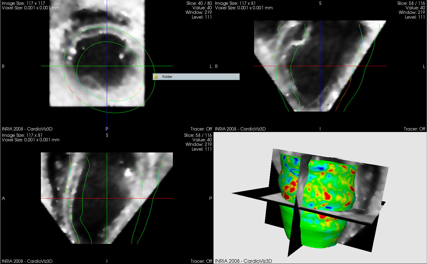
Figure 3: Example of 4D Visualization in 4D displacement and strain in the cardiac.
Thus, we propose to extensively test the hypothesis that measures of 4D displacement and strain, obtained with our method using 4D ultrasound has clinically equivalent results compared to the gold standards of MR-based methods and sonomicrometry, and the hypothesis that 4D methods have advantages in performance for strain estimation over current commercialized 2D methods. This step is critical and will be a significant advance to make this new technology available for use in clinical research and practice. As we point out in Section B, we are going through the same path that MR tagging and 2D speckle tracking techniques went through in their early stages of development. Our hope is that once this step is finished, it will open the door and allow this technology to be applied more broadly to clinically important and compelling research applications (e.g., dyssynchrony) , similar to how MR tagging (and other MR phase tracking methods) and 2D speckle tracking methods are being used today. Thus, the three specific aims of our proposed efforts are summarized below.
Specific Aim 1 Experimental Validation: Quantify errors of displacement and strain in optical flow on 4D ultrasound with respect to similar measures obtained by sonomicrometry in in vivo canine (dog) hearts.
Specific Aim 2 Clinical Validation: Test the hypothesis that optical flow measures computed from 4D ultrasound can yield clinically equivalent results on healthy volunteers (normal hearts) and diseased hearts compared to similar measures obtained by state-of-the-art MR based methods.
Specific Aim 3 Clinical Evaluation: Test the hypothesis that optical flow on 4D ultrasound has specific quantitative advantages in terms of performance when compared to 2D based methods of strain estimation.
Support
NIH 1R01HL086578-01A2
Publications
Qi Duan,Andrew F Laine,Elsa Angelini, Assessment of fast anisotropic diffusion and scan conversion of real-time three-dimensional spherical ultrasound data for visual quality and spatial accuracy Proceedings of SPIE, Medical Imaging, Vol. 5373, pp. 331-342 , 2004
Qi Duan,Elsa Angelini,Ting Song,Andrew F Laine, Fast interpolation algorithms for three-dimensional real-time cardiac ultrasound Proceedings of the 25th Annual International Conference of the IEEE Engineering in Medicine and Biology Society (EMBS), pp. 1192-1195, 2003
Qi Duan,Elsa Angelini,Shunichi Homma,Andrew F Laine, Tracking Endocardium Using Optical Flow along Iso-Value Curve 28th Annual International Conference IEEE Engineering in Medicine and Biology Society (EMBS), pp. 707-710, New York City, USA., 2006
D. Moses,M. Srichai,V. Pai,Qi Duan,Andrew F Laine, Semi-Automatic Ventricular Border Segmentation Package Based on Multi-Phase Levelset Segmentation International Society for Magnetic Resonance in Medicine (ISMRM), 14th Scientific Meeting & Exhibition, 2006
Q. Duan,S. Herz,C. Ingrassia,O. Gerard,K. Costa,J. Holmes,Shunichi Homma,Andrew F Laine,Elsa Angelini, Dynamic Cardiac Information From Optical Flow Using Four Dimensional Ultrasound Proceedings of 27th Annual International Conference IEEE Engineering in Medicine and Biology Society (EMBS), pp. 4465-4468, 2005
A. Patel,P. Robson,C. McKenzie,D. Sodickson,Andrew F Laine,Qi Duan, Rapid Reference-Free Noise Reduction for Parallel MR Images Using a Principal Component Technique in Combination with Adaptive Dyadic Wavelet-Based Denoising ISMRM 16th Scientific Meeting & Exhibition, Toronto, Canada, 2008
V. Pai,Qi Duan,Andrew F Laine, Real-Time Myocardial Segmentation in MRI ISMRM 16th Scientific Meeting & Exhibition, Toronto, Canada, 2008
V. Pai,Jian Chen,Andrew F Laine,Qi Duan, Dyadic Wavelet-Based Image Noise Suppression and Enhancement in High-Speed Cardiac MR Parallel Acquisition ISMRM 16th Scientific Meeting & Exhibition, Toronto, Canada, 2008
Qi Duan,Elsa Angelini,Shunichi Homma,Andrew F Laine, Real-Time Segmentation Of 4D Ultrasound By Active Geometric Functions Proceeding of 5th IEEE International Symposium on Biomedical Imaging (ISBI): From Nano to Macro, pp. 233-236, 2008
G. Shechter,L. Gutiérrez,D. Stanton,L. Zagorchev,D. Elgort,Qi Duan,Andrew F Laine, Augmenting CT Cardiac Roadmaps with Segmented Streaming Ultrasound Proceedings of SPIE Medical Imaging, Vol. 6509, pp. 65090V-1 - 65090V-11, 2007
O. Gerard,K. Costa,J. Holmes,Shunichi Homma,Andrew F Laine,Qi Duan,Elsa Angelini, Cardiac Motion Analysis Based on Optical Flow on Real-Time Three-Dimensional Ultrasound Data Recent Advances in Diagnostic and Therapeutic Ultrasound Imaging for Medical Applications, Suri et al Eds., Artech House Press, 2006
M. Schrijver,Q. Duan,R. Kemkers,Qi Duan,Andrew F Laine,Gert Schoonenberg, Adaptive spatial-temporal filtering applied to x-ray fluoroscopy angiography Proceedings of SPIE, Medical Imaging, Vol. 5744, pp. 870-878, 2005
S.L. Herz,O. Gerard,P. Allain,C.M. Ingrassia,K.D. Costa,J.W. Holmes,Shunichi Homma,Qi Duan,Elsa Angelini, Tracking of LV Endocardial Surface on Real-Time Three-Dimensional Ultrasound with Optical Flow Proceedings of the 3rd International Conference on Functional Imaging and Modeling of the Heart (FIMH), LNCS Vol. 3504, pp. 434-445, 2005
O. Gerard,,Qi Duan,Elsa Angelini,Shunichi Homma,Andrew F Laine, Comparing Optical-Flow Based Methods for Quantification of Myocardial Deformations on RT3D Ultrasound Proceedings of IEEE 2006 International Symposium on Biomedical Imaging (ISBI); From Nano to Macro, pp. 173-176, 2006
S.L. Herz,C.M. Ingrassia,O. Gerard,K.D. Costa,J.W. Holmes,Andrew F Laine,Qi Duan,Elsa Angelini, Evaluation of Optical Flow Algorithms for Tracking Endocardial Surfaces on Three-Dimensional Ultrasound Data Proceedings of SPIE, Medical Imaging, Vol. 5750, pp. 159-169, 2005
Qi Duan,Elsa Angelini,S. Herz,C. Ingrassia,K. Costa,J. Holmes,Shunichi Homma,Andrew F Laine, Region-Based Endocardium Tracking on Real-Time Three-Dimensional Ultrasound Ultrasound in Medicine and Biology, Vol. 35, No. 2, pp. 256-265, 2008
O. Gerard,K. Costa,J. Holmes,Shunichi Homma,Andrew F Laine, Cardiac Motion Analysis Based on Optical Flow of Real-Time 3-D Ultrasound Data Page 227-246. Moli-Suri, Editors, Artech House, Boston MA, 2008
Qi Duan,Andrew F Laine,Elsa Angelini, Chapter 20: Tracking Endocardium Using Optical Flow along Iso-Value Curve Principles and Recent Advances in Medical Imaging and Image Analysis, Dhawan et al Eds., World Scientific Publishing Company, pp. 337-366 , 2008
Qi Duan,Shunichi Homma,Elsa Angelini,Andrew F Laine,Auranuch Lorsakul, Coronary Occlusion Detection with 4D Optical Flow Based Strain Estimation on 4D Ultrasound Proceedings of Functional Imaging and Modeling of the Heart (FIMH), pp. 211-219, Nice, France, 2009
Yrjö Häme,Viktor Gamarnik,Katherine M. Parker,Jeffrey W. Holmes,Andrew F Laine,, Level set-based tracking of the endocardium without a shape prior from 3D ultrasound images International Symposium on Biomedical Imaging, 2012